
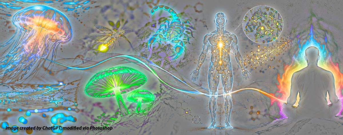
Question for you biologists: What do Virginia opossums, bark scorpions, Mexican free-tailed bats, and a species of Javan orchid have in common?
Answer: They are members of a vast population of organisms that exhibit something called biofluorescence.

You may recall that I introduced this new series of posts at the end of September with a discussion of a phenomenon called bioluminescence, which occurs through a chemical reaction that produces light energy within an organism’s body. Part 2 of this series will shine a light (!) on a similar phenomenon, biofluorescence.
Biofluorescence differs from bioluminescence in that the organisms that exhibit it do not generate light on their own. Instead, they absorb light (or radiation) from an external source, and then emit it at a different wavelength. Also unlike bioluminescence, biofluorescence is not the product of a chemical reaction; it requires an external light source and an internal fluorescent biomolecular substance (which we’ll discuss in a moment; don’t be alarmed by the fancy words as the concept isn’t that complicated).
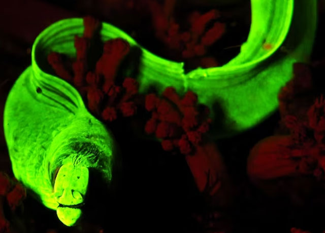
Photo credit: Maarten De Brauwer, Senior Research Scientist at Southern Cross University
In addition, biofluorescence is generally undetectable by the unaided human eye. But, as we shall see (!), the the equipment and conditions that allow us to detect glowing organisms are relatively simple and easy to obtain. But, first, let’s explore the mechanism by which biofluorescence occurs.
How does biofluorescence work?
Excellent question…thanks for asking! Do you want the long answer [1] or the short one? Never mind; I’m going to provide the latter, partially because I don’t fully understand the details of the former, but mainly because those details aren’t all that important for most of us (if, however, you’re doing post-graduate work in the field of Fluorescence Microscopy, feel free to check out the footnote linked above).
Biofluorescence is the absorption of light by an organism at one wavelength followed by its reemission at a different wavelength. A molecule within the organism’s cells absorbs short-wavelength, high-energy light such as ultraviolet (UV) or blue light, then releases some of that energy as longer-wavelength light such as yellow or red. While that may sound as though the organism is simply reflecting the incoming light, that isn’t what’s happening. The light is being absorbed, its energy is being changed, and a different wavelength of light is emitted.
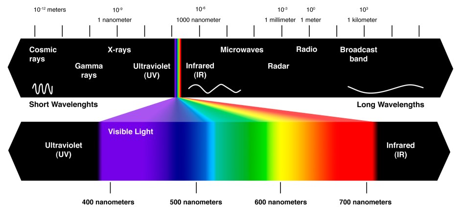
The following illustration shows the range of wavelengths, from incredibly short cosmic rays to very long (1,000 miles or more) radio waves. It shows that visible light occupies a pretty tiny sliver of the overall spectrum (click to see a larger version).
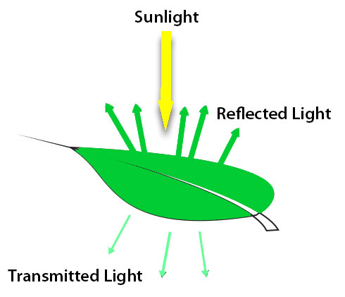
Living organisms have within their cellular structure a molecular component called a chromophore. The chromophore absorbs some wavelengths of incoming light, and allows others to be reflected or transmitted, which gives the organisms their color. Chlorophyll is an example; it’s a collection of molecules in which chromophores capture light for use in photosynthesis. It strongly absorbs light in the blue and red wavelengths; the green wavelength is less strongly absorbed and is reflected or transmitted [2], thus giving many plants that color.
But this is not biofluorescence.
Biofluorescence occurs when some of the energy of absorbed light is stored, a tiny fraction of it is lost as it is converted to heat, and the remainder is then emitted at a different (lower-energy) wavelength. This process takes place in an incredibly short period of time, one that is measured in picoseconds . A picosecond is one-trillionth of a second, slightly faster than the time it takes for me to forget the name of a person I just met. [3]
This process involves another molecular component called a fluorophore. The term excitation describes what happens when a fluorophore absorbs light energy and enters a higher-energy state. At the risk of getting too detailed, remember that light is composed of photons. When a photon of the appropriate wavelength is absorbed by a fluorophore, it excites the molecule. As the molecule returns to a lower-energy state, it may release some of that energy as a photon of light. The emitted light generally has a longer wavelength than the absorbed light, and therefore a different color. (You might want to refer to the illustration above to see how color varies with wavelength.) The wavelength of the emitted light depends on several factors, including the fluorophore’s chemical structure and its molecular surroundings.
As an interesting side note, fluorophores in non-living tissue will fluoresce under the appropriate wavelength of light, meaning that even ancient museum samples can be valuable for research into biofluorescence.
So, that’s a 50,000′ view of the science behind biofluorescence. If you’re still awake at this point — good work! We can now move to the really fun stuff, starting with…
Where can we find biofluorescence?
Everywhere. Or, to be more accurate…what organisms display biolfluorescence? A whole bunch. Or, to be more accurate…thousands upon thousands of species of fish, birds, fungi, arthropods, plants, amphibians, reptiles, and mammals (including humans!). In fact, according to the authors of this article, biofluorescence may well be the default condition for all mammals.
Now, just because something exhibits biolfluorescence doesn’t mean that it goes around constantly glowing…at least not as far as human viewership is concerned. The light that’s emitted is generally so weak that it’s not visible to the unaided eye. Also, the cells that fluoresce may be internal to the organism. We’ll talk more about how biofluorescence can be viewed, but let’s look at a brief list of species whose fluorescence might surprise you. That list includes:
| Species | Color of fluorescence |
| Mediterranean house gecko | Neon green |
| Millipede | Greenish-blue |
| Amaneti cokeri mushroom | Blue |
| Ball python | Orange |
| Green bell pepper | Red |
| Flying squirrel | Pink |
| Porcupine (quills) | Blue-green |
| Platypus | Purple-green |
| Cowries (seashell; some) | Red |
| Emperor penguin | Blue |
| Salmon-crested cockatoo | Gold-orange |
| Springhare | Multiple (see photo below) |
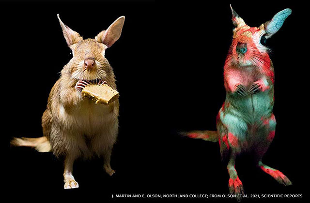
A precise number fluorescent organisms is impossible to state with any certainty, but here is one estimate by category:
- Invertebrates, fungi, plants — thousands of species
- Birds — hundreds of species
- Amphibians and reptiles — hundreds of species
- Mammals — almost all species, and there are almost 7,000 recognized species to date
The National Wildlife Foundation provides another snapshot of fluorescent organisms found in North America. Keep in mind that the species count differs in some instances from the preceding estimates, demonstrating the challenge of testing the millions of species of living organisms in the world.
| Animal Group | Confirmed Species | Glow Color(s) | North American Examples |
| Reef-building corals | ~850 | Green, orange, red, cyan | None native |
| Fish | 459+ | Green, red | Many reef & freshwater species |
| Amphibians | All 32 species tested; likely universal | Green, blue, orange | Frogs, toads, salamanders |
| Birds | 421+ | Blue, green, pink | Owls, nighthawks, others |
| Mammals | 125 | Pink, cyan, green | Opossum, flying squirrel, porcupine, bats |
| Reptiles | 7+ gecko, 80+ chameleon | Blue-green | Mediterranean house gecko |
| Arthropods (non-scorpion) | Widespread; 1000s estimated | Variable | Millipedes, crab spiders, many moths |
| Scorpions | All 2500 known species | Blue-green, cyan, neon green | Striped bark scorpion |
What are the purposes of biofluorescence?
As far as I can tell, the answers to this question are varied, but they have one thing in common: they’re simply educated guesses. Still, they’re based on logical assumptions that are often based on direct observation, and scientists continue to seek confirmation that those assumptions are correct. Here are the most common explanations.
- Intra-species communication — The fluorescence of some organisms is visible only to other members of the species. This has been observed specifically in some deep sea fish whose predators can’t detect the emitted fluorescence. This could provide a basis for attracing a mate (without attracting a predator), or for warning others about the presence of danger.
- Camouflage — The fluorescence of some species — again, mostly in marine organisms — helps them blend in with their surroundings.
- Luring of prey — Blue or ultraviolet light attracts insects (see also: electric bug zappers), and so species such as Venus flytraps and, again, some species of fish use biofluorescence to attract meals.
- Warning to predators — The use of aposematic Aposematism is the advertising by an animal or plant, whether terrestrial or aquatic, to potential predators that it is not worth attacking or eating. use of biofluorescence has been observed or hypothesized in a variety of organisms. For example, some theorize that the fluorescence of a porcupine’s quills are a signal to predators of the danger in attempting to attack it.
- Incidental chemistry — This is a fun one. According to this article via the National Wildlife Federation, the biofluorescence in mammals such as bats, platypuses, and flying squirrels has no apparent use, and it might simply be a matter of having certain naturally occurring compounds in their fur. Keratin, in particular, fluoresces, and the structural framework for all mammalian fur is keratin.
What about biofluorscence in humans?
It’s logical to assume that the first four factors listed above are not relevant to humans, since we can’t perceive UV with our unaided eyesight, at least not in the normal course of life. (I suspect that the exception would be if your “normal course of life” includes spending significant time in carnival fun houses and night clubs. You know who you are.) So that leaves us with the “incidental chemistry” explanation for why certain parts of our anatomy are biofluorescent when exposed to UV light. Those parts include:
- Teeth — Certain components of tooth enamel glow blue-white under a black light.
- Fingernails and toenails — Portions of these glow a subtle white or blue under a black light.
- Skin — Certain proteins found in skin react to UV exposure, as do some lipids (fat) on the surface of skin.
In addition, various components of the human body contain endogenous fluorophores, which are naturally fluorescent compounds that react to UV or infrared light, but that reaction is not detectable at the surface level. Examples include some coenzymes, A coenzyme is an organic, non-protein molecule that enhances the function of an enzyme. An enzyme is a protein that helps biochemical reactions occur. collagen, and porphyrins. Porphyrins are specialized molecules characterized by their ability to capture metal ions, and they play crucial roles in various biological and chemical processes. For example, porphyrin complexes called hemoglobin are found in the red blood cells of all mammals. In plants, porphyrins combine with magnesium to form chlorophyll. The human body also secretes fluids that fluoresce, but I’ll let you use your imagination or do your own research on that if you’re curious.
There’s an entire body of science called photobiology that studies the effects of light on living organisms. The details of those studies particularly with respect to their application to medical diagnoses and treatments, are fascinating but well beyond the scope of this post.
How can we see biofluorescence?
The most common way to detect it is by shining a UV-emitting black light (aka UV flashlight) [4] on the subject. The UV rays will excite (see above) the organism’s fluorophores, and result in an emission of visible light of varying colors, as shown in the examples in the table above. This process is sometimes referred to as induced fluorescence.
It’s also possible to photograph instances of biofluorescence. The technique is called Ultraviolet-induced fluorescence (UVIF). UVIF is best done with a DSLR or mirrorless camera, but it’s also possible with a cellphone equipped with a high quality camera. [5]
Looking ahead…
We’re two-thirds of the way through this series, having first covered the phenomenon of bioluminescence and now, biofluorescence. If you’ve made it this far, you have my undying admiration, because the subject matter is dense, and my attempts to elaborate on it may have been equally challenging.
The final chapter will deal with something called Ultraweak Photon Emission, and it may well be the most interesting of the three phenomena. I hope you’ll check back to find out what that’s all about.
Footnotes
[1] I find it a bit intimidating that it takes a 9,000 word essay to explain Basic Concepts in Fluorescence, but here we are. I really did try to read the essay, but references to Jablonski energy diagrams, Planck’s constant, femtoseconds A femtosecond is one-quadrillionth of a second, the approximate time it takes me to say ‘yes’ to another order of nachos. , and fluorophores, not to mention the well-studied probe fluorescein isothiocyanate (FITC) only served to make me want to take a nap. And yet…that fluorophore thing will actually pop up again, and can’t be ignored, even for a basic explanation of the phenomenon of fluorescence. So, you can either read the linked essay, or [Return] for a more…um…accessible description.
[2] The term transmitted in this context merits some clarification. Think about looking down at a green leaf on a plant on a sunny day. The surface of the leaf appears green because light with that wavelength is being reflected. But if you look up at the underside of the leaf, backlit by the sun, the green color is the result of transmitted wavelengths that moved through the leaf. Perhaps this simple graphic will help explain the process:
[Return]
[3] This will likely never appear as a question in your next bar trivia game, but the conversion of invisible short wavelength rays such as ultraviolet to visible longer wavelength light is called the Stoke’s shift. It’s named for the Irish physicist, Sir George Stokes, who first described the phenomenon. Sir George was, among other things, a baronet, which sounds suspiciously like a woodwind or other musical instrument but is less interestingly a British hereditary title of nobility. I mention this only because this footnote would be too short without it. And possibly less informative. But a burning question remains, and that’s why the shift in Stoke’s shift is not capitalized. I’ve done extensive research on this question via a single Google query, and it turns out that due to the immutable rules of eponym, findings such as this merit the capitalization of only the finder’s name. See also, Doppler effect. There. Now the footnote has met its quota. [Return]
[4] The purpose of a black light within the context of biofluorescence is to project ultraviolet rays onto an organism in order to excite its fluorophores (if, indeed, they are present), causing them to emit visible light of various colors.
Despite their usefulness in detecting biofluorescence, the most common uses for UV flashlights seem to be: (1) to identify the presence of scorpions, either inside or outside a home; (2) to view fluorescing rock samples (which is not biofluorescence given that, as far as we can tell, rocks are not alive); and (3) to identify pet- and human-related stains on floors and bedding in defiance of the idea that ignorance is bliss.
Regardless of how one intends to use a UV flashlight, one needs to be aware of certain safety considerations. Ultraviolet light can be categorized as UV-A (the lowest energy, and thus the safest), UV-B (higher energy, and the type that causes sunburn), and UV-C (the highest energy, used for disinfectant purposes, and capable of quickly and severely damaging skin and eyes). To ensure that your flashlight is the safest (you still never want to shine it in your or someone else’s eyes), look for one with a wavelength of 365nm (recommended) or 395nm (acceptable, with some drawbacks); both emit only UV-A.
365nm UV light is less visible to the human eye and thus doesn’t produce glare that competes with emitted biofluorescence. It also delivers more energy per photon than 395nm, and thus induces a stronger fluorescent reaction from organisms. On the other hand, 395nm UV light is partially visible to the human eye in the form of a purple or violet glow and can overpower certain colors of biofluorescence. 395nm UV is more commonly used for entertainment purposes — e.g., in a nightclub or for viewing glow-in-the-dark artwork — where a focused beam is not desirable.
395nm UV flashlights tend to be less expensive; you might think of them more as a general purpose UV flashlight than one specifically for viewing biofluorescence. [Return]
[5] Here’s how to go about taking photos of biofluorescence (as well as fluorescence emitted by minerals, ceramics, and other non-organic materials).
You’ll first want to find or place your subject in a setting that’s as dark as possible. Any ambient light will diminish the fluorescence.
You’ll then need a good camera capable of a long exposure — typically a DSLR or mirrorless camera — and a tripod, ideally with a remote shutter release to avoid camera shake. Here are the suggested settings for your camera:
- Lens — A 100mm macro lens is ideal, but not required
- Mode — Manual, with manual focus; try shining a white light on the subject using autofocus, then switch to manual to lock in the focus setting
- Aperture — f/2.8 to f/22 to let in as much light as possible
- ISO — start at 1000, but depending on the results, you change to a lower ISO to reduce noise in the image, or a higher one if you want a brighter picture
- Shutter speed — 1 to 20 seconds or more for closeups, e.g., fungi or macro photography; you might need to experiment to get the best results
- White balance — Custom or daylight/cloudy (around 5000K–5500K); do not use the Auto white balance setting
- UV light source — 365 nm UV light source is recommended (see Footnote 4 for more information); you won’t need a special filter on your lens if you do.
Once you have your camera setting fixed, illuminate the subject with your UV flashlight and trip the shutter; again, for best results, use a remote shutter release to eliminate camera shake.
Don’t have a fancy DSLR? Not a problem…you can also take photos of UVIF with an iPhone or Android phone that has a camera with adjustable settings. I’m not going to get into that here, but feel free to google the topic and you’ll find simple instructions for the necessary settings, depending on the model of your phone. [Return]
Bibliography
My research for this post took me to around forty different websites — not counting several dozen inquiries to Wikipedia — along with queries to A.I. (primarily ChatGPT and Google’s Gemini) as I attempted to understand the science and technology associated with a topic that I knew almost nothing about. What follows is not a true bibliography, but it’s a partial list of the online resources that I relied on to varying degrees while composing the article. I’ve linked to many of them in the body of the text, but I decided to provide a more comprehensive list in case any readers want to get deeper into the subject. Links are listed in no particular order.
- ScienceNews — Backyard explorers discovered 15 new examples of glowing life
- National Geographic — Pink squirrels and green sharks—why do so many animals ‘glow’ in UV light?
- iNaturalist — Salmon-crested cockatoo observations
- National Library of Medicine — Aposematism: balancing salience and camouflage
- National Library of Medicine — The Covert World of Fish Biofluorescence: A Phylogenetically Widespread and Phenotypically Variable Phenomenon
- National Wildlife Federation — New in Wildlife Science: Fluorescent Mammals & Penguin Power Naps
- National Wildlife Federation — Glowing in the Dark: The Secret UV Lightshow in Your Backyard
- The Royal Society Publishing — All-a-glow: spectral characteristics confirm widespread fluorescence for mammals
- The Royal Society Publishing — Glandular biofluorescence in fire salamanders (Salamandra salamandra): first evidence and ecological implications
- Molecular Expressions — Basic Concepts in Fluorescence
- HunterLab — The Difference Between Biofluorescence and Bioluminescence
- ABC News (Australia) — The eerie glow of biofluorescence — and how it can help us conserve marine life
- Forestry and Land Scotland — Biofluorescence in the rainforest
- Finding Fluorescence — What is biofluorescence?
- Understanding Animal Research — Biofluorescence: mammals that glow in the dark
- Exotic Esoterica — Ultraviolet Fluorescence in Plants: Light in the Fast Lane
- Spectroscopy — Unveiling the Mysteries of Scorpion Fluorescence: Insights from Ultraviolet Excitation
- Science Over a Cuppa — AUTOFLUORESCENCE: WHEN TISSUES LIGHT UP UNDER YOUR MICROSCOPE FOR NO APPARENT REASON…
- BioOne Digital Library — Fluorescence in Fireflies (Coleoptera: Lampyridae): Using Sentinel Prey to Investigate a Possible Aposematic Signal
- Interesting Engineering — This is How Biofluorescence Works in Nature
- Reddit — Best blacklight for bioluminescence
- American Society of Mammologists — The Mammal Diversity Database
- ScienceDirect — Photon Excitation
- ScienceDirect — Coenzyme
- AAT Bioquest — What are the differences between a fluorophore and a chromophore?
- EMR-TEK — 365 vs 395 Light: What’s the Difference?
- Petapixel — How to Capture Ultraviolet-Induced Fluorescence (UVIF) Photos
- EBSCO Information Services — Research Starter: Chlorophyll
- EBSCO Information Services — Research Starter: Porphyrin
Discover more from The Fire Ant Gazette
Subscribe to get the latest posts sent to your email.